| 1. Praestigia duffeyi Millidge, 1954 |
|
| 2. Praestigia kulczynskii Eskov, 1979 |
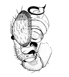
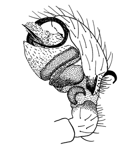
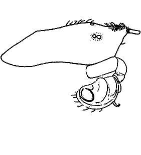
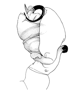
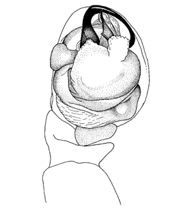
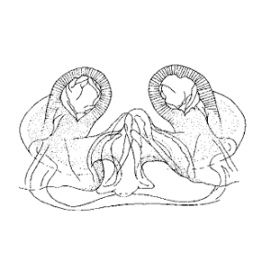
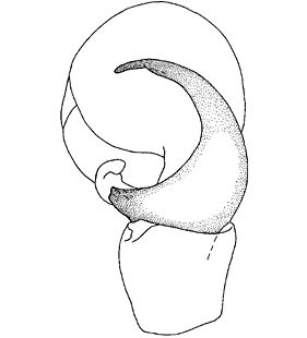
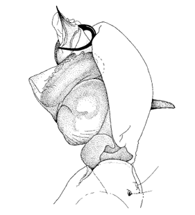
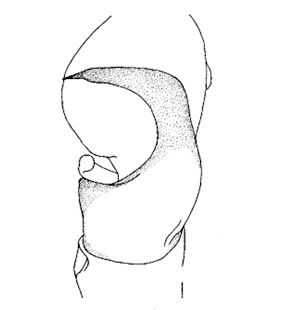
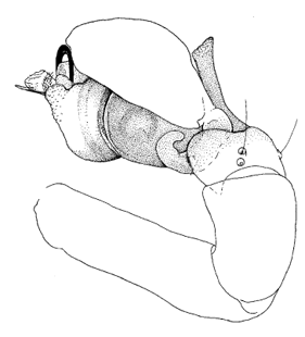
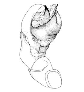
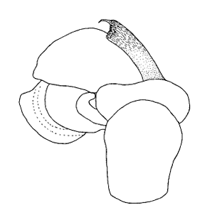
  Pedipalpus, lateral Pedipalpus, lateral
(Paquin & Dupérré 2003)
|
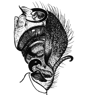
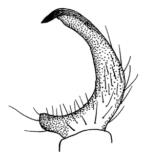
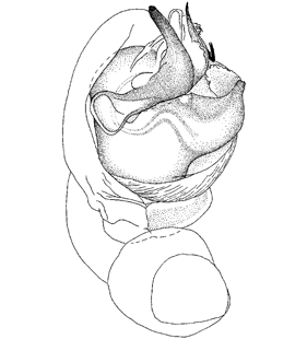
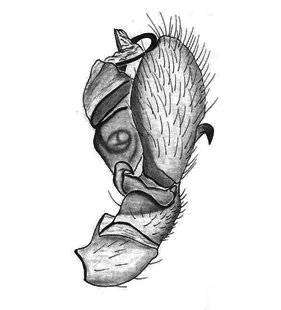
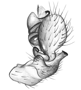
 Pedipalpus, retrolateral
(Marusik et al. 2008) |
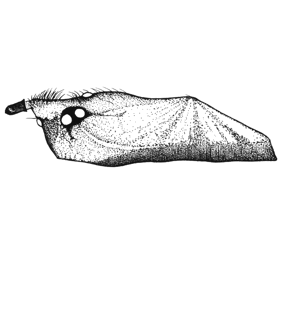
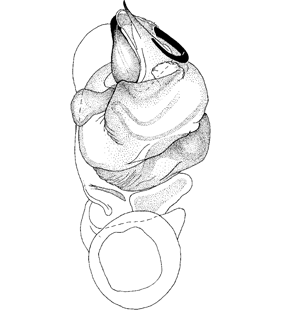
 Pedipalpus, prolateral
(Marusik et al. 2008) |
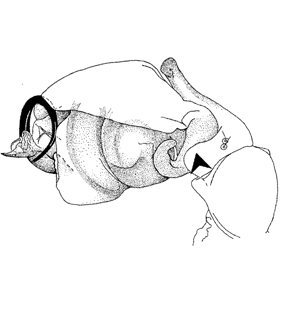
 Pedipalpus, ventral
(Marusik et al. 2008) |
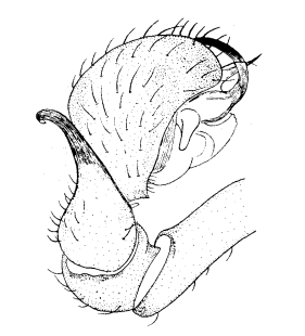
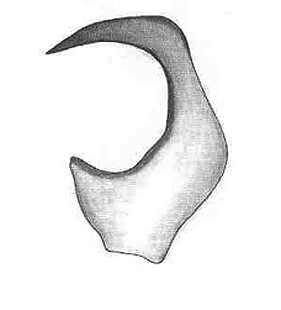 Pedipalpentibia
(Paquin & Dupérré 2003) |
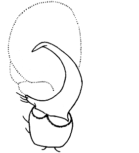
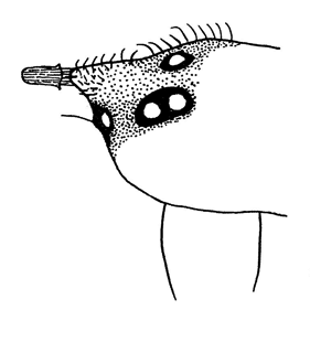
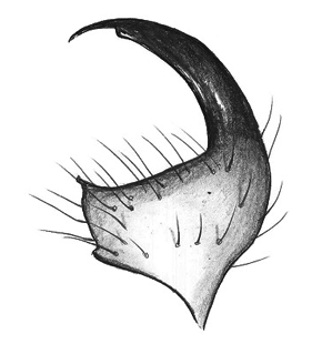
 Tibialapophyse, dorsal
(Marusik et al. 2008) |
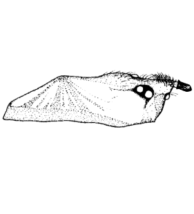
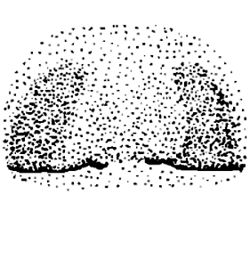
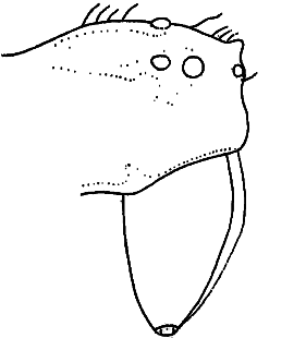
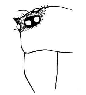
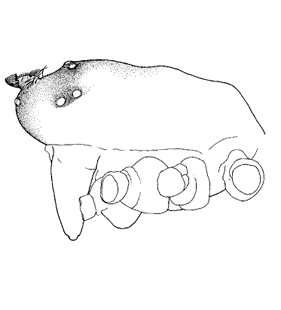
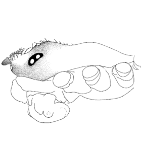
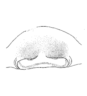
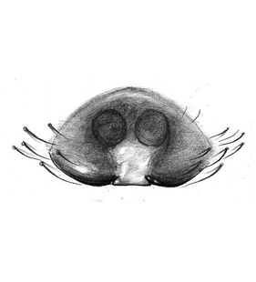
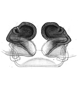
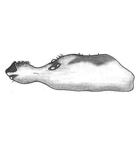 Prosoma, lateral
(Paquin & Dupérré 2003) |
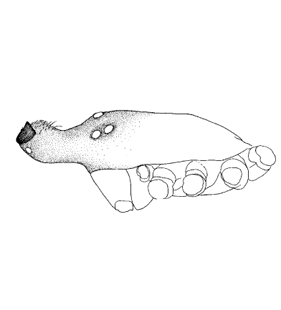 Prosoma, lateral
(Marusik et al. 2008) |
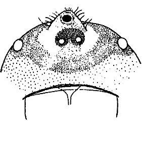
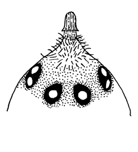
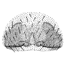
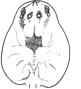
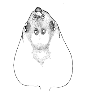
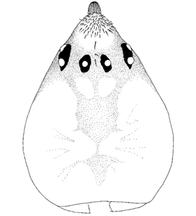
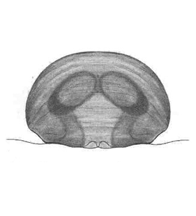
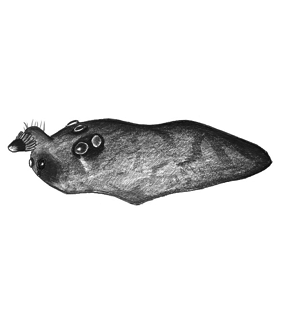
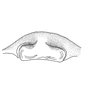
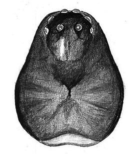
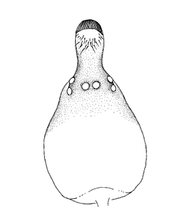 Prosoma, dorsal
(Marusik et al. 2008) |
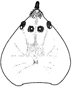
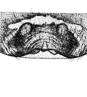
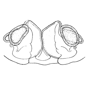
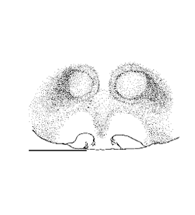
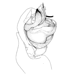
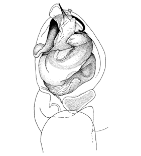
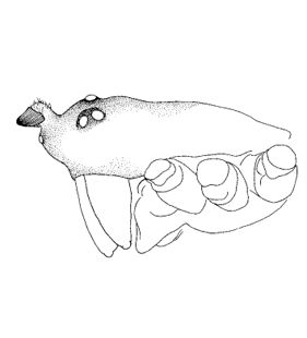
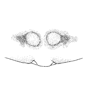
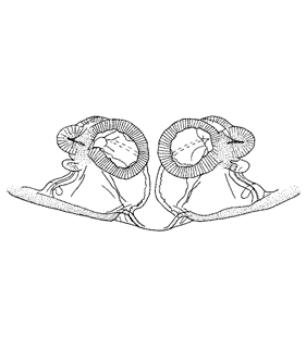
  Epigyne Epigyne
(Paquin & Dupérré 2003)
|
 Epigyne, caudal
(Marusik et al. 2008) |
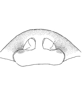
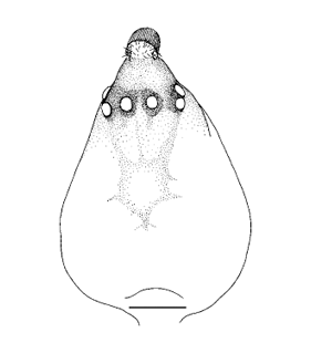
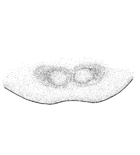 Epigyne, ventral
(Marusik et al. 2008) |
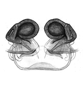
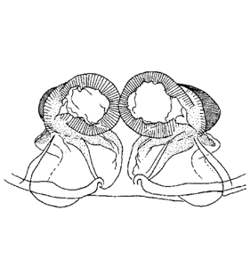 Vulva, ventral
(Marusik et al. 2008) |
|
|
|
| 3. Praestigia makarovae Marusik, Gnelitsa & Koponen, 2008 |
|
| 4. Praestigia pini (Holm, 1950) |
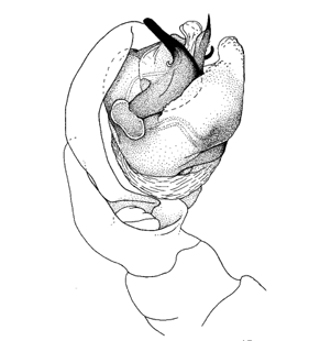
 Pedipalpus, retrolateral
(Marusik et al. 2008) |
 Pedipalpus, prolateral
(Marusik et al. 2008) |
 Pedipalpus, retrolateral
(Løvbrekke unpubl.) |
 Pedipalpentibia, dorsal
(Løvbrekke unpubl.) |
 Tibialapophyse, caudal
(Marusik et al. 2008) |
 Tibialapophyse, dorsal
(Marusik et al. 2008) |
 Prosoma, lateral
(Marusik et al. 2008) |
 Prosoma, dorsal
(Marusik et al. 2008) |
 Prosoma, lateral
(Løvbrekke unpubl.) |
 Epigyne, ventral
(Løvbrekke unpubl.) |
 Epigyne, caudal
(Marusik et al. 2008) |
 Epigyne, ventral
(Marusik et al. 2008) |
 Vulva, ventral
(Løvbrekke unpubl.) |
 Vulva, dorsal
(Løvbrekke unpubl.) |
 Vulva, ventral
(Marusik et al. 2008) |
 Prosoma, dorsal
(Løvbrekke unpubl.) |
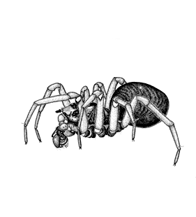
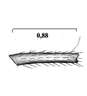
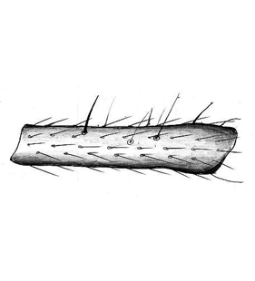 Tibia I
(Løvbrekke unpubl.) |
 Metatarsus I
(Løvbrekke unpubl.) |
| |
|
|
|
| 5. Praestigia uralensis Marusik, Gnelitsa & Koponen, 2008 |
|